Medical imaging can help diagnose bone tumors
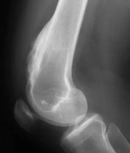
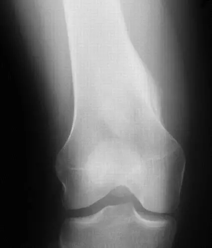
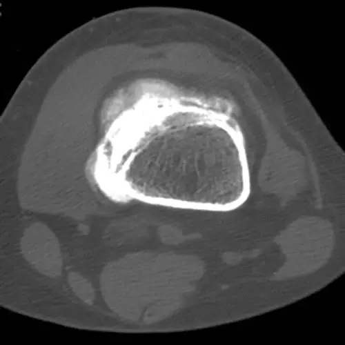
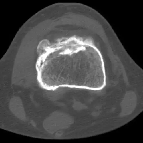
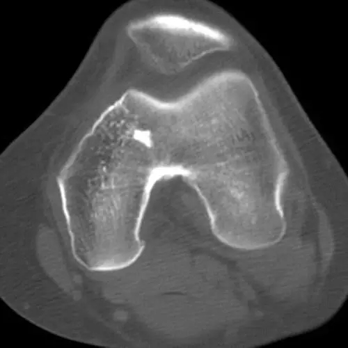
A 20-year-old woman has painful knee pain that lasts for several months. She denied major trauma or previous surgery. Radiographs were obtained and then CT and MRI examinations were performed. The patient is then referred to a specialist clinic for diagnosis and definitive treatment. The positive lateral radiograph of the knee joint (Figure 1-2). On the lateral radiograph, a proliferative bone-like bulge dominated by mature bone is formed along the anterior medial aspect of the distal femur. The lesion does not appear to encroach on the medullary cavity. Focal sclerosing lesions can be seen in the distal lesion of the metaphysis. The fat on the palate moves forward and the edges are clear. In the AP view, the cortex is slightly expanded in the medial direction. There are no fractures. figure 1 figure 2 Expert Discussion (Dr. Richardson): Morphologically, most of this lesion looks like mature bone tissue. In my experience, more than 95% of this result will be considered heterotopic ossification, and in the remaining cases, the same is true for the bone series of tumors. The following are my differential diagnosis: (1) heterotopic ossification, (2) osteochondroma, (3) osteoma, (4) osteoid osteoma, (5) osteoblastoma, (6) macular degeneration Tumor and (7) osteosarcoma. Osteochondroma is a possibility, but tumors can often see dysplasia of some tissue structures around the periphery. The lesion appears to be obtained after the bone has matured because the shape and proportion of the femur are normal. The distal femur is not a good site for osteoma or osteoblastoma, and there is no characteristic pain in the lesion (nocturnal pain relieved by aspirin), so the possibility of osteoid osteoma is also reduced. However, I still feel the need Make a cross-section to find the nest. Limeratosis (Melorheostosis) is a strange shape. I have seen a crescent-shaped change. Therefore, I usually don't mention it in most differential diagnoses. However, it Can be similar to heterotopic ossification, osteoma, or even osteosarcoma, so in some special cases I put him in the differential diagnosis. Most types of osteosarcoma usually appear to be more invasive and visually impactful in general; in this case, I seriously consider locking the disease in a highly differentiated periosteal sarcoma. Radiographs are very bad in showing soft tissue extension and I expect to be found in osteosarcoma; this gives us a reason to get CT or MRI. The small lesion at the distal end is the bone island. image 3 Figure 4 Figure 5 Endoscopic Discectomy Instruments Endoscopic Discectomy Instruments,Endoscopy Discetomy System,Transforaminal Surgical System,Orthopaedics Diskoscope Set ZHEJIANG SHENDASIAO MEDICAL INSTRUMENT CO.,LTD. , https://www.sdsmedtools.com