Characterization of Quantum Dots and EGFR Antibody Conjugates and Their Tumor Cell Binding Properties
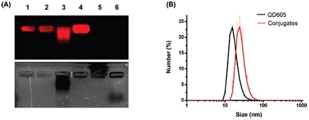
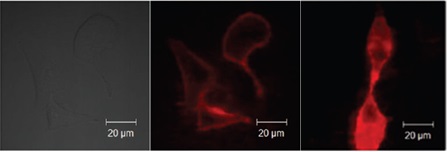
Abstract: Cetuximab/Erbitux is the first anti-tumor monoclonal antibody targeting EGFR. It is a conjugate of a new generation of fluorescent probes, quantum dots, for high expression of EGFR. In vitro and in vivo imaging of tumor cells. Epidermal growth factor receptor (EGFR) is a tumor marker and is highly expressed in many types of tumors such as breast cancer, lung cancer, prostate cancer and rectal cancer. It is an important prognostic indicator. Cetuximab/Erbitux is the first anti-tumor monoclonal antibody to target EGFR. Compared with traditional organic fluorescent dyes, quantum dots have excellent photochemical properties such as high fluorescence intensity and strong fluorescence stability, and are a new generation of fluorescent labeling probes. Labeling EGFR antibodies with quantum dots can be used for in vitro and in vivo imaging studies of tumor cells with high expression of EGFR. The Rita Song team in Korea applied three different coupling strategies to obtain biologically functional quantum dot-Erbitux conjugates for tumor-specific imaging. The use of PEG-coated quantum dots and two long-chain diisofunctional linkers sulfo-LC-SPDP and sulfo-SMCC successfully coupled quantum dots with Erbitux (Fig. 1). The dissociation constant of the conjugate and EGFR was 0.61±0.28 nM. The conjugate was characterized by ultraviolet-visible spectrometer, fluorescence spectrophotometer, agarose gel electrophoresis, BCA protein concentration determination and dynamic light scattering. The conjugate was observed to bind to living cells and colocalize with transferrin by laser confocal microscopy (Fig. 2). The above experimental results provide a new research method for imaging tumor cells with high expression of EGFR, which can provide important information for related drug treatment and prognosis analysis. Figure 1 Agarose gel electrophoresis (A) and dynamic light scattering (B) confirm the coupling of quantum dots with Erbitux The upper graph in Figure A shows the fluorescence imaging of the quantum dot QD605, and the lower panel shows the Coomassie blue staining after the corresponding electrophoresis. The lane samples were: 1. Quantum dot QD605 (control); 2. Quantum dot activated by sulfo-LC-SPDP; 3. Quantum dot-Erbitux conjugate (coupled by LC-SPDP and SMCC linker) ; 4. Quantum Dots - Erbitux conjugate (coupled by EDC); 5. Erbitux (EGFR antibody); 6. Erbitux activated by sulfo-SMCC. Electrophoresis conditions: 1% agarose gel / 0.5x TBE buffer (pH 8.5), 50 Vcm -1 , 40 min. Figure 2 In vitro cell imaging of quantum dots-Erbitux conjugates Left: bright field imaging of A549 cells cultured in vitro; medium: quantum dot-Erbitux conjugate was incubated with A549 cells for 20 min; right: quantum dot-Erbitux conjugate was incubated with A549 cells for 1 h. Source of the document: Lee J, Choi Y, Kim K, Hong S, Park HY, Lee T, Cheon GJ, Song R. Characterization and cancer cell specific binding properties of anti-EGFR antibody conjugated quantum dots. Bioconjug Chem. 2010;21(5): 940-6. Outdoor Solar Camera,Surveillance Camera Wireless,Home Security Cctv Camera,Solar Powered Camera Shenzhen Zuomi Technology Co., Ltd. , https://www.leftriceptz.com