
Application of fluid shear system in biomedical research
Gastric Lavage Training,Vital Sign Simulation,Nasal Cavity Structure Model,Anatomy Nose Nasal Cavity Model YinChuan EXin Medical Instrument Co.,Ltd , https://www.exmedmodel.com
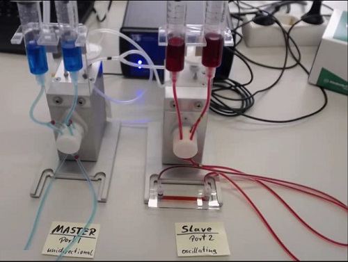
A fluid shear system is a device that produces a liquid flow by applying air pressure within a fluid-filled accumulator. The device circulates liquid by using a special switching mode to produce a constant one-way flow. This makes it an ideal setting for applying defined shear stresses in long-term cell culture. Continuous and pulsating laminar flow and oscillating flow can be simulated using a fluid shear pump system. Thereby simulating the fluid flow environment of blood vessels, lymphatic vessels and other tissues under physiological conditions, and then carrying out related cell research work. Guangzhou Keshi Special Science Instrument Co., Ltd. has long been committed to fluid-related experimental research and technical services. The fluid shearing system is briefly introduced in biomedical research as follows. 
The following is a brief summary of several common applications of fluid shear systems.
I. Long-term cell culture under flow
Principle: In vivo, endothelial cells develop and differentiate under shear stress conditions. When starting to use endothelial cells for cell-based testing, you should consider the possible effects of this mechanical force on cell morphology and physiology. In order to place the cells in a more physiological, in vivo state, they are cultured for several hours to several weeks under flow, producing more relevant results. In other words, flow regulation is critical for any type of study using cells with physiologically potential flow conditions.
Application Examples <br> Study the effects of shear stress on endothelial cell physiology through various experimental readings, such as immunofluorescence, Western blot, qPCR and FACS
Prepare the cell layer for subsequent functional assays such as rolling and adhesion or transfer assays 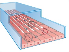
Flow characteristics Continuous laminar flow, non-uniform flow, oscillating flow (required for disturbance flow simulation), pulsating flow Experiment duration Hours, several weeks Recommended pump Ibidi fluid pump system Recommended μ-Slides μ-Slide I Luer,
μ-Slide VI0.4
μ-Slide Y shape
Second, rolling and adhesion analysis
Principles <br> In rolling and adhesion assays, white blood cells and/or platelets are perfused on the surface with a protein coating or cell layer. Their adhesion to the surface and interaction with the surface can be analyzed under various conditions (for example, after gene knockdown or drug treatment).
Applications
1 Study the adhesion of platelets and leukocytes to endothelial cells or matrix proteins 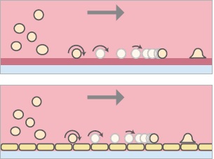
Flow characteristics Continuous laminar flow Experiment duration Minutes to hours Recommended pump Ibidi pump system syringe pump peristaltic pump Recommended μ-Slides μ-Slide I Luer μ-Slide VI 0 .4
μ-Slide Y shape
Third, the definition of liquid exchange
Principles <br> Channel slides and pumps are used to provide cells with precisely defined amounts of media and/or any supplements of interest.
Applications
1 Define the exchange of cell culture media
2 Living Ca2+ - Imaging
3 Live cell imaging of drug-stimulated adherent cells
4 Live cell imaging of cell staining 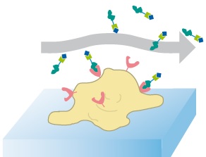
Flow characteristics Short laminar flow Experiment duration Minutes to hours Recommended pump Manual liquid delivery, syringe pump, peristaltic pump Recommended μ-Slides μ-Slide VI0 .4
μ-Slide I Luer
Fourth, 3D cell culture
Principle <br> Cells are cultured in a 3D gel matrix. The interstitial stream is applied directly to the gel to provide nutrients O2 and CO2 to the cells to maintain optimal conditions.
Application Examples <br> 3D culture of single cells, spheroids or organoids in the gel matrix (eg, with hepatocytes, fibroblasts, myocytes, kidney cells and stem cells) 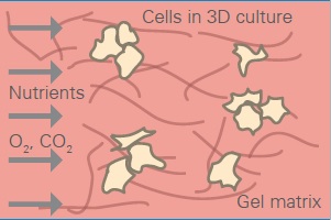
Flow characteristics Gap flow is very low Experiment duration Hours up to several weeks Recommended pump Ibidi fluid pump system Recommended μ-Slides μ-Slide VI0 .4
μ-Slide I Luer
V. Cellular electrical impedance sensor (ECIS) flow analysis
The cell electrical impedance sensor (ECIS) is a platform for measuring the morphological and physiological changes in the adherent cell layer by impedance. Living cells can be measured directly in the container without any interference and do not require any staining. The ECIS flow array enables researchers to combine any type of flow measurement with parallel impedance measurement. 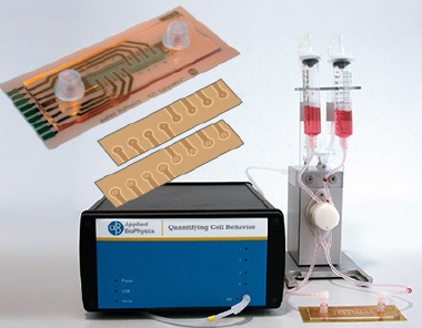
Six references
1. Schulz C, et al. (2009) Novel Methods for Assessment of Platelet and Leukocyte Function Under Flow - Application of Epifluorescence and Two-Photon Microscopy in a Small Volume Flow Chamber Model. Open Biol J 2(1): 130–136 .